Stub Procedure
- We collected pollen from our three flower specimens: Tulip (Tulipa), Wisteria (Disambiguation), and Snapdragon (Antirrhinum). We did this by using tools such as scalpels to extract the pollen from the anthers of the flowers.
- Next, we placed the pollen of the flowers on three separate portions of a stub for the SEM microscope. We did this by using a paint brush to dust the pollen on on its specific portion of the stub.
Light Microscope Pollen Photos
This is a microscope photo of pollen from a tulip blossom.
This is a photo from the light microscope of an anther from a wisteria blossom.
This is a photo from the light microscope of a pollen sample from snap dragon.
SEM Work
This is a photo of snap dragon from a distance.
This is a photo of piece of snap dragon pollen being measured in the SEM.
This is a close up of one piece of snap dragon pollen.
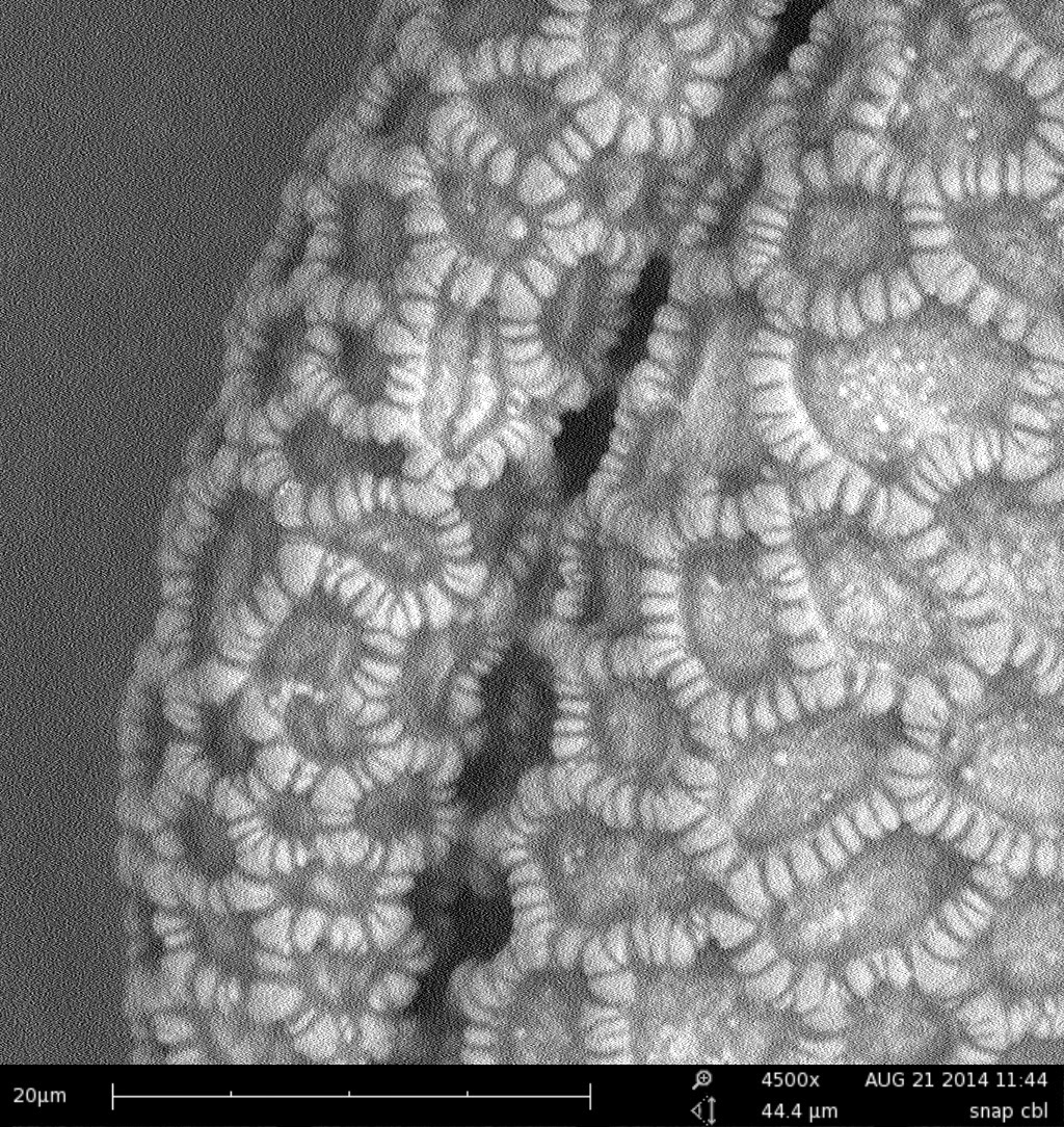
This is an incredible close up of snap dragon pollen. As you can see, the texture of the pollen is intricate and there's tons of details.
This is an overview of tulip pollen.
This is a photo of a bit of tulip pollen being measured by the SEM.
This is a close up to see the texture the tulip pollen in the SEM. As you can see, both the shape and texture of the tulip pollen is very different from snap dragon pollen.
This is an overview of the anther from a wisteria blossom.
This is a photo of an anther from a wisteria blossom being measured in the SEM.
Lieca microscope
- We scraped pollen from each flower on to the tray for examination.
- We then examined the pollen under the microscope:


No comments:
Post a Comment